

Acne vulgaris is not a sudden overnight event. It is a gradual process inside the pilosebaceous unit. Many people notice a red, firm bump only when inflammation is already visible, yet a microcomedone can form deeper in the follicle long before it can be seen.
From non-inflammatory comedones to red papules, pus-filled pustules and deeper nodules or cysts, recognising each stage helps you care for the skin more carefully. Avoiding forceful squeezing and seeking a dermatologist when needed are important for skin health and for lowering longer-term scar risk.
Key points:Acne develops as excess sebum and abnormal keratin build-up, microbial growth that triggers immune inflammation, neutrophil accumulation with pus formation, and—if the follicle wall ruptures—deeper tissue injury. Non-inflammatory comedones can worsen into papules and pustules after pressure or ongoing irritation. Once the follicle wall breaks in the dermis, painful nodules or cysts may form. Do not force open persistently red or deep lumps; have a clinician assess them in person.
Where does acne start? Tiny changes in the pilosebaceous unit
Acne mainly appears where sebaceous glands are denser, such as the T-zone, cheeks, chest and back. Keratinocytes in the follicle normally shed with sebum and exit to the surface. When androgen-driven sebum increases and follicular keratinocytes become stickier and shed abnormally, keratin mixes with sebum into a microcomedone that is hard to see at first.
Microcomedones are the shared starting point of acne lesions. As sebum and keratin keep packing the follicle, the follicular opening stretches and visible comedones appear. For everyday triggers, see why acne keeps coming back.
Stage 1: How do comedones form? Closed whiteheads vs open blackheads
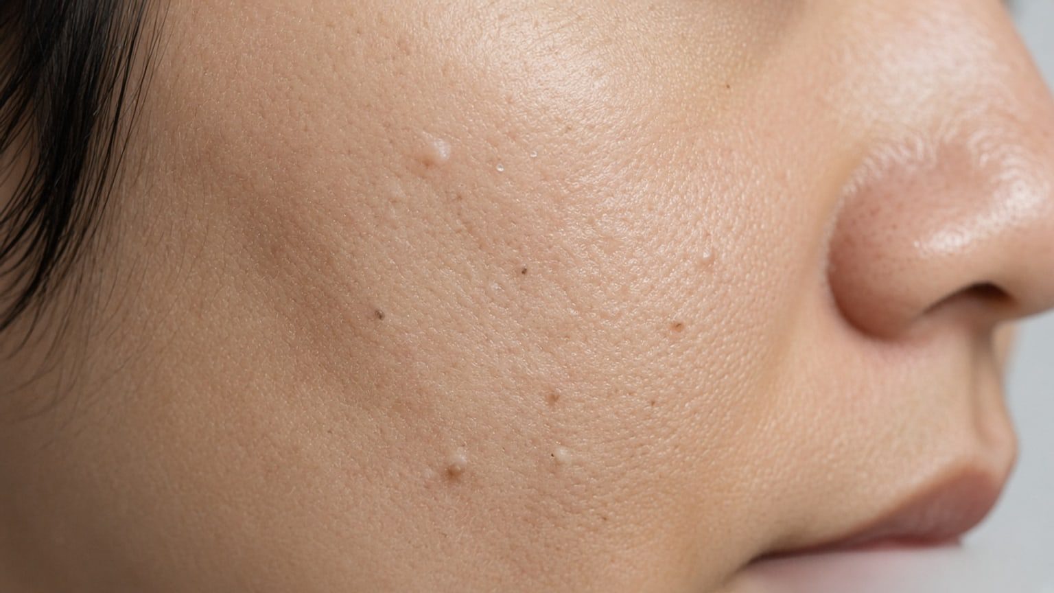
Closed comedones (whiteheads)
When excess sebum and keratin collect inside the follicle while a thin keratin layer still covers the opening, a closed comedone—or whitehead—forms. It looks like a skin-coloured or whitish tiny bump without a clear open pore. It may feel finely grainy but is not red or tender. Because the outlet is closed, the anaerobic environment favours microbial overgrowth and raises the chance of later inflamed acne.
Open comedones (blackheads)
When the follicular opening widens as the plug expands, the tip is exposed to air and an open comedone—or blackhead—forms. The dark colour is not simply dirt. Melanin in keratinocytes within the sebum oxidises on contact with air, and light refraction adds to the dark appearance.
Comedones are non-inflammatory lesions; surrounding tissue has not yet mounted a clear immune response. Focus on gentle cleansing and basic moisturising. Avoid aggressive extraction tools or fingernail digging, which can break the barrier and turn a quiet comedone into red inflammation.
Stage 2: Why do comedones turn red? How papules form and look
Cutibacterium acnes growth and free fatty acid irritation
The follicle normally hosts microbes, including anaerobic Cutibacterium acnes. When a comedone fully occludes the follicle and sebum keeps flowing, an oxygen-poor, nutrient-rich niche forms. The bacteria produce lipase that splits triglycerides into free fatty acids, which chemically irritate the follicle wall.
Immune-cell infiltration and papule formation
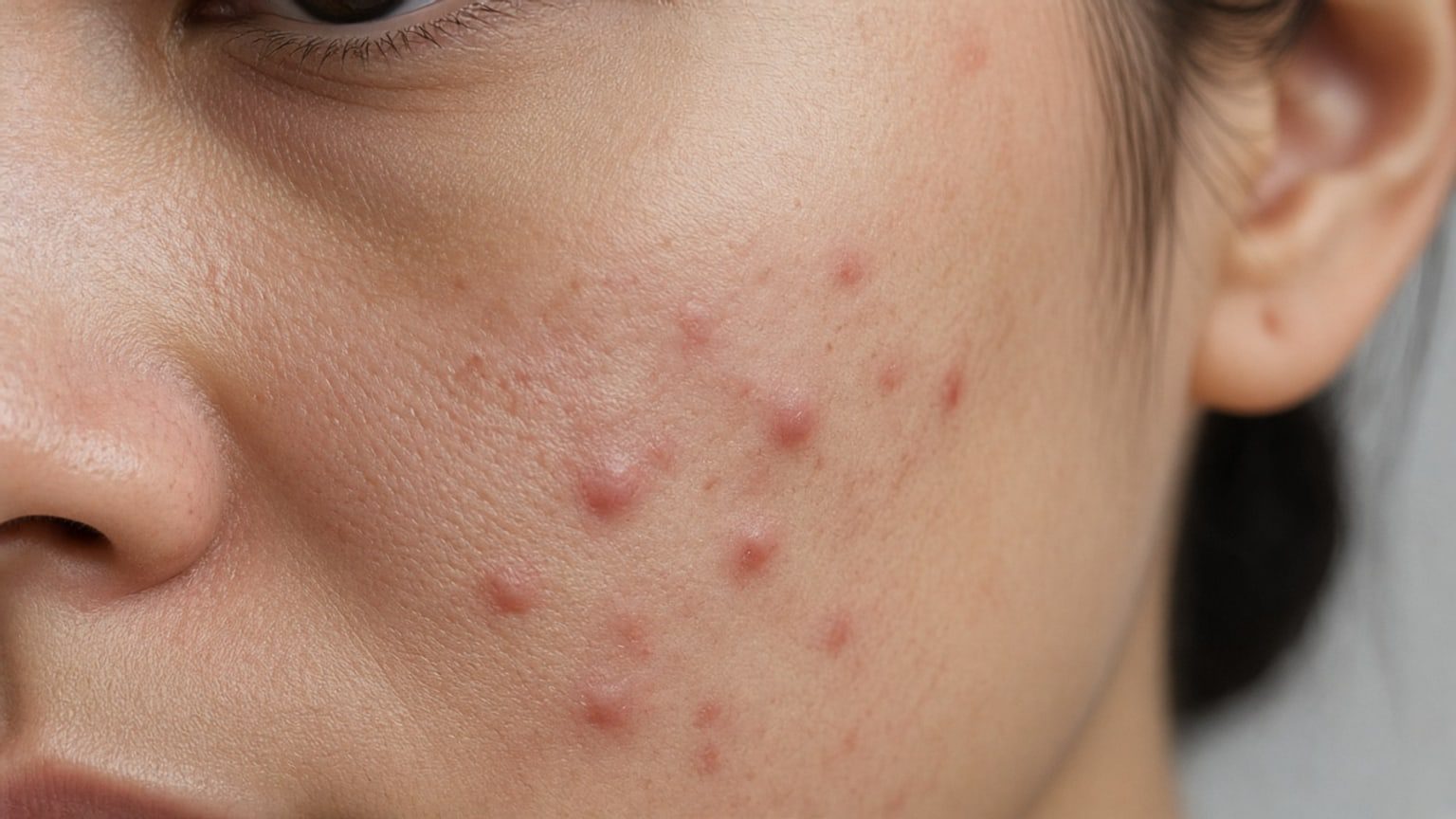
Free fatty acids and bacterial products push follicular epithelium to release pro-inflammatory cytokines and recruit white blood cells, especially neutrophils. Nearby vessels dilate and tissue swells, forming a red papule usually under 0.5 cm.
Papules feel firmer and mildly tender. They mainly reflect congestion and cellular infiltrate without organised superficial pus. Trying to squeeze often yields only watery fluid and may drive inflammation deeper into the dermis.
Stage 3: Why do red spots fill with pus? Pustule features and change
Neutrophil accumulation and pus formation
As immune cells and Cutibacterium acnes interact intensely, many neutrophils enter and engulf bacteria. Dying white cells, bacterial debris, shed keratin and liquefied sebum mix into yellowish or greyish sticky pus.
Pustule appearance and fragile structure
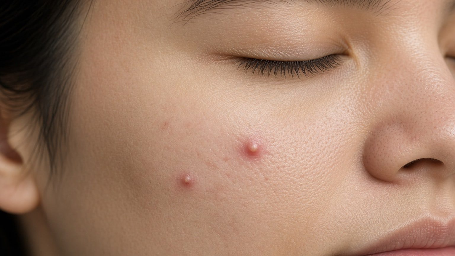
When pus rises toward the follicular tip, a visible yellow-white head forms on a red base—a pustule. The overlying epidermis is thinned by inflammation and easily disrupted.
Pustules often throb or sting on touch. Even when the head looks shallow, do not pierce with unsterile tools or pinch hard with nails. Poor technique can rupture the weakened follicle wall downward or sideways and spread infection into deeper soft tissue.
Stage 4: When inflammation reaches the dermis—nodules vs cysts
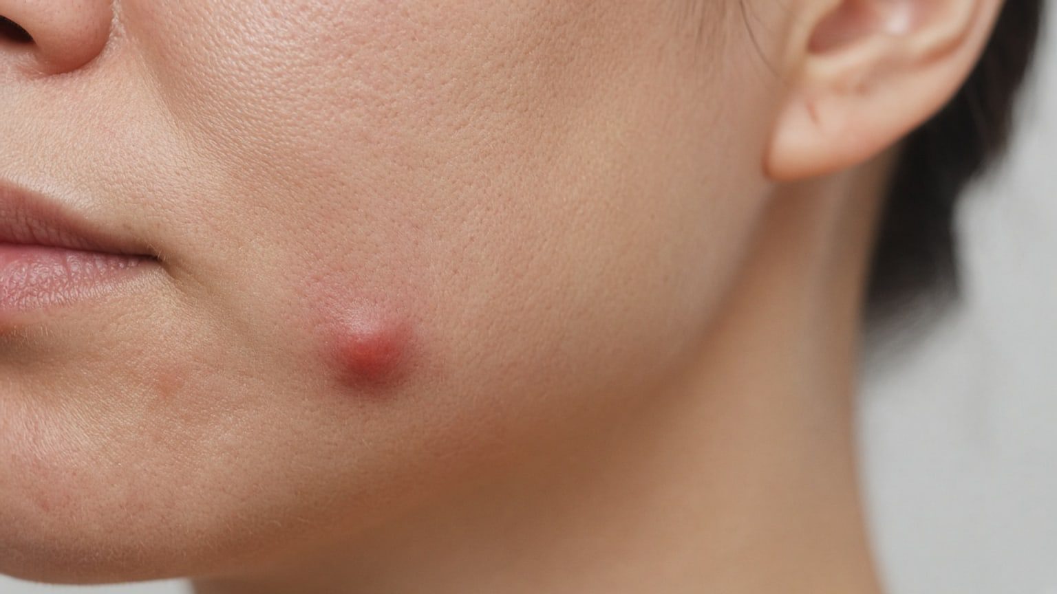
Follicle-wall rupture and deep granulomatous inflammation
If internal pressure is high or strong external squeezing occurs, the inflamed follicle wall can rupture deep in the dermis. Irritating sebum, keratin fragments, dead bacteria and inflammatory mediators spill into sterile dermis and subcutaneous fat, triggering intense foreign-body granulomatous inflammation—stage four.
Nodule features
Nodules are usually deep inflammatory lumps larger than 0.5 cm. The surface may look only mildly raised and dusky red, yet they feel hard and broad under the skin, with deep aching and marked tenderness. They are solid masses of dense inflammatory and granulation tissue, not superficial pus that can be expressed.
Cyst features and how they differ from nodules
Cysts form when deeper inflammation liquefies and leaves a cavity filled with abundant pus and necrotic debris. They are often larger, softer and fluctuant. Severe lesions may interconnect under the skin as sinus tracts, heal slowly and readily destroy dermal collagen, leaving atrophic or hypertrophic scars that are hard to reverse.
How can you quickly compare the four acne stages and care points?
To recognise the current stage more clearly in daily life, compare non-inflammatory comedones through deep nodules and cysts by lesion type, mechanism, typical look and care principles:
| Stage | Lesion type | Main mechanism | Typical look and feel | Care principles |
|---|---|---|---|---|
| Stage 1: comedones | Closed (whiteheads) / open (blackheads) | Increased sebum plus abnormal keratinisation plugs the follicular opening | Flat or slightly raised white dots, or a dark keratin plug at an open pore; no heat, redness or pain | Gentle cleansing; avoid highly comedogenic products; do not dig or squeeze |
| Stage 2: papules | Inflamed red bumps | Occlusion lets Cutibacterium acnes grow and break down sebum, driving local immune infiltrate | Red raised papule under 0.5 cm, mild surrounding redness, slight tenderness | Pause harsh exfoliation and irritating care; do not squeeze by hand; ask a clinician about anti-inflammatory topical options |
| Stage 3: pustules | Superficial pustular lesions | Many neutrophils gather, and debris forms pus | Yellow-white visible head on a clearly red base; stinging on touch | Keep the area clean and dry; avoid piercing that spreads infection; if it opens on its own, blot gently with clean cotton |
| Stage 4: nodules & cysts | Deep hard inflammatory lumps / cysts | Follicle-wall rupture spills contents into deep dermis with widespread granulomatous inflammation | Nodules hard and deeply painful; cysts larger and fluctuant, sometimes interconnected | No external squeezing; high scar risk—seek prompt medical care |
Can you squeeze acne yourself? Follicle damage from improper pressure
Seeing a red bump or yellow-white head often triggers the urge to squeeze. Force from fingers or unsterile tools pushes in more than one direction: only part of the contents may exit, while irritating inflammatory material is driven deeper into the dermis.
The most common harm is rupturing an already fragile follicle wall. A superficial papule or pustule can rapidly become a deep nodule or cyst, prolong the course for weeks, and damage elastic fibres and collagen—leaving ice-pick or rolling scars and post-inflammatory pigment that may last months to years.
Build a habit of not touching, picking or squeezing. For more home-care principles, see how to improve acne.
When should you seek professional care? Visit timing and preparation
Warning signs that warrant earlier review
Acne is not only a teenage skin change. Repeated inflammation can affect skin structure over time. Seek professional advice if you have deep, very tender nodules on the face or body; dense pustules that are spreading; clear pits or pigment after lesions settle; or if non-prescription anti-acne products used for several weeks leave the skin no better—or more red, peeling and stinging.
Individual assessment in dermatology clinic
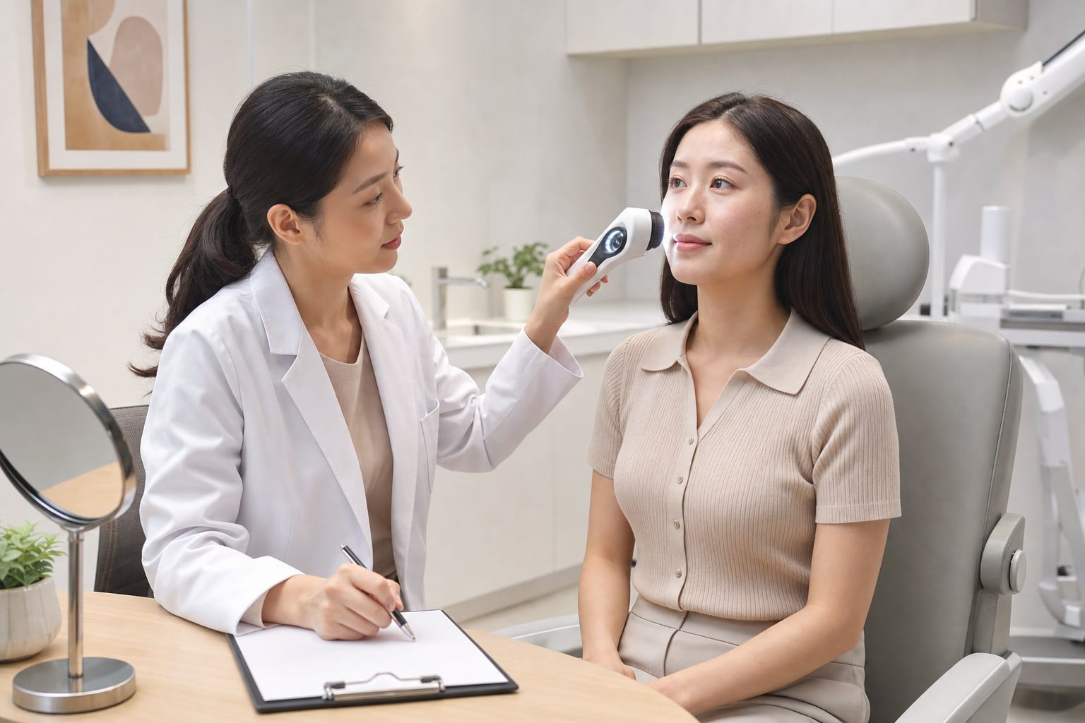
A dermatologist reviews which formation stage your lesions are in, their distribution and your skin type, then plans care with your routine and product habits in mind. Read acne treatment options and how to prepare for an acne visit. In central Taiwan you can arrange assessment at the dermatology clinic, or browse the health library for more skin guidance.
Both are non-inflammatory comedones. Closed comedones (whiteheads) have an opening covered by keratin, so sebum has little air contact and appear as tiny skin-coloured or white dots. Open comedones (blackheads) have a widened opening; the plug darkens mainly from melanin oxidation after air exposure. Do not force extraction by hand; ask a dermatologist if clearing is needed.
No. Many comedones settle or stay non-inflammatory with gentle care. Progression to papules, pustules or deep cysts is more likely when sebum is high, Cutibacterium acnes proliferates, or friction and squeezing add injury. Early gentle habits help interrupt the inflammatory pathway.
Papules are early inflammation with congestion and infiltrate, not organised superficial pus. Forceful squeezing often yields nothing useful and can push mediators deeper, rupture the follicle wall and leave nodules or depressed scars. Keep the area clean and ask a clinician about suitable topical care.
Blot fluid gently with clean cotton. Do not keep squeezing around the wound. After cleaning, saline compresses or a clean breathable covering may help; avoid unsterile tools. Seek care if redness, heat or drainage continues so infection risk can be assessed.
Nodules are usually deep, firm inflammatory lumps over 0.5 cm with clear tenderness. Cysts are deeper, often softer cavities with liquid pus or necrotic debris and a fluctuant feel. Both involve deep dermis or subcutis; ordinary leave-on products rarely reach them, and squeezing risks extensive tissue damage and atrophic scars—medical care is required.
Seek dermatology help for deep red painful hard lumps; numerous inflamed papules and pustules over a wide area; clear pits, raised scars or dark marks as spots settle; or when non-prescription products used for a period bring no improvement and cause more redness or sting. Early stage-based assessment helps lower long-term scar risk.
Doctor’s note: identify the stage—do not squeeze inflamed lesions blindly
From microcomedones and red papules to deep nodules, each stage has a different mechanism and care logic. Many stubborn lumps and scars follow forceful squeezing that ruptures the follicle wall rather than the natural course alone. If pustules, deep hard painful lumps or home care that is not improving appear, avoid picking and have a dermatologist assess the stage and plan individualised care to reduce tissue injury and lasting scar risk.
This article provides general disease education and is not a medicine advertisement or product recommendation. It cannot replace an in-person examination or individual treatment advice.
Dermatology medical team

Dr. Tseng