

Moles and spots are not the same thing
A mole is a cluster of melanocytes forming a structure that can be flat or raised, sitting anywhere from the epidermis to the deep dermis. A spot is usually a colour change caused by pigment deposition, without necessarily any thickness of its own.
That is why they are approached differently. For a mole, the nature and depth need confirming first. For a spot, what matters is which type it is and how deep the pigment sits. Treating different things the same way makes both the result and the risk hard to control.
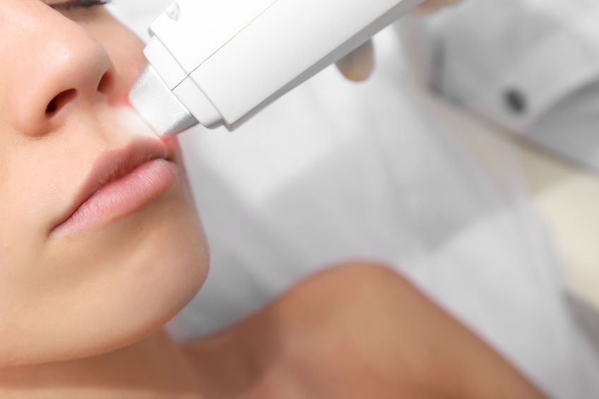
What kinds of spots are there?
They are all called spots, but the depth and cause differ a great deal, and so do the suitable approach and the number of sessions needed.
| Type | Appearance and location | Pigment depth | Approach |
|---|---|---|---|
| Sun spots | Brown patches with a clear border on sun-exposed areas such as the face and backs of the hands | Superficial | Relatively straightforward, but sun protection afterwards decides how soon they return |
| Freckles | Small, clustered light brown spots that darken with sun exposure | Superficial | Treatable, but long-term sun protection is needed to limit recurrence |
| Melasma | Blurred brown-grey patches, usually on the cheeks and forehead | Deeper and uneven | Energy needs to be conservative; too high and it can darken. Often combined with other treatment |
| Cafe-au-lait macules | Light brown patches with a fairly clear border, often present from childhood | Superficial to mid | Treatable, but recurrence is relatively common, so expectations are set beforehand |
| Hori nevus | Symmetrical grey-blue dots over both cheekbones | Dermal | Needs several sessions with longer intervals |
| Nevus of Ota | Blue-grey pigmentation around one eye and the temple | Dermal | Needs several sessions and a plan set by the doctor |
Melasma and birthmark-type pigmentation particularly need careful assessment. Using the wrong energy on a misidentified lesion can make the pigment darker rather than lighter.
Which moles need assessing first?
Most moles are benign, but a small number can change. ABCDE is the commonly used self-check prompt. If any of these apply, it is better to have a dermatology specialist examine it than to book a laser session.
| Warning sign | What it means |
|---|---|
| A for Asymmetry | Fold the mole in half and the two halves look clearly different |
| B for Border | The edge is jagged, blurred or extends outwards |
| C for Colour | Shades vary within the same mole, or red, white or blue appears |
| D for Diameter | Larger than about 6 mm, or noticeably bigger than before |
| E for Evolving | The shape, colour or height changes, or it starts to itch, bleed or ulcerate |
New pigmented lesions under a nail, on the sole, palm or a mucous membrane are also worth having examined. Lesions in those locations are harder to judge on your own.
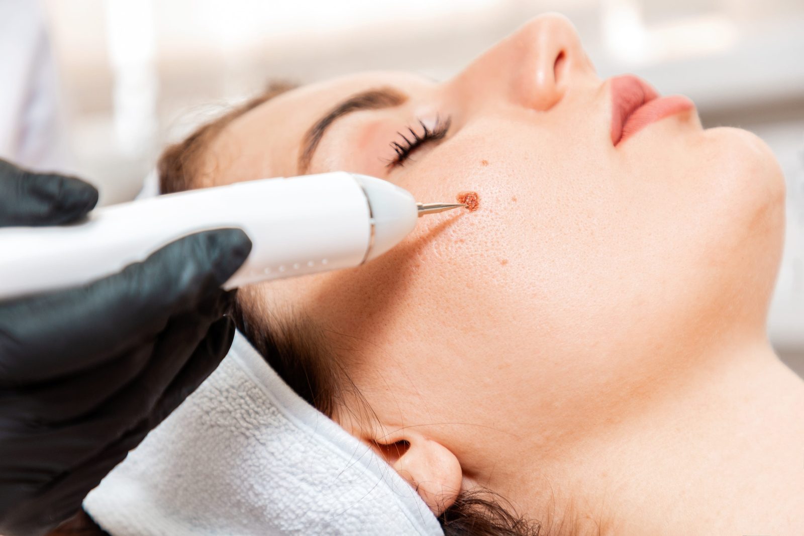
When is laser not the right approach?
A lesion that is suspicious or of uncertain nature is excised surgically and sent for pathology, not lasered. Laser vaporises the tissue, so there is no specimen left to examine, and the chance to confirm the diagnosis is lost.
It is also worth discussing timing with your doctor if the area is inflamed or has a wound, you have had heavy sun exposure or are currently sunburnt, you have a keloid tendency or a history of abnormal scarring, you are taking photosensitising medication or an oral retinoid, or you are pregnant or breastfeeding.
What are the options?
What is actually done depends on the nature, depth, location and size of the lesion, as decided by the doctor.
| Approach | Worth discussing for | Points to note |
|---|---|---|
| Laser | Benign, superficial moles and pigmented spots | Nothing remains to send for pathology, so it is used only where the lesion is clearly benign |
| Surgical excision with pathology | Suspicious or uncertain lesions, or deeper and larger moles | Confirms the diagnosis at the same time; sutures are removed at a follow-up and a linear scar remains |
| Electrocautery or shave removal | Some raised benign lesions | Suitability is judged by the doctor; aftercare is similar to laser |
| Topical treatment and sun protection | Supporting and maintaining results for superficial pigmentation | Limited on its own, usually used alongside a course of treatment |
Responses vary from person to person. This table is to help you understand the options before your visit and is not a guarantee of results.
What does a session involve?
For laser treatment of a benign lesion, a session is usually completed within about 40 minutes, most of which is waiting for the topical anaesthetic.
- Assessment: the doctor examines the type, depth and location of the lesion and confirms whether laser is suitable or a biopsy is needed first.
- Topical anaesthetic: about 30 minutes.
- Laser: roughly one to five minutes, depending on the number and size of lesions.
- Aftercare: a hydrocolloid dressing or ointment is provided as appropriate, with instructions.
- Follow-up: deeper lesions may need several sessions, usually reviewed four to six weeks apart.
How do you look after the area afterwards?
Aftercare directly affects the chance of pigmentation and scarring, and this part is down to you.
- Sun protection: avoid direct sun for a week and use a high-factor sunscreen plus physical cover when going out.
- Keep the wound clean and dry, following your instructions. Where cleaning is needed, wipe gently with saline rather than rubbing.
- Do not pick the scab. Let it come away on its own; picking makes pigmentation or scarring more likely.
- Use the dressing or ointment as directed and do not substitute an unidentified product.
- If redness worsens, there is pus or marked pain, return to have it assessed for infection.
Brief redness, bruising or stinging usually settles within a few days. The chance of pigmentation and scarring depends on your skin, the site and the aftercare.

A note from the dermatology team
Removing a mole or a spot looks like a cosmetic question, but the first step is really a safety one: confirming whether this mole is benign and which type this patch of pigmentation is. Get the order right and the approach and the expectations that follow both make sense.
If a mole has recently changed shape, colour or size, or a new pigmented lesion appears under a nail or on the sole, it is worth having a dermatology specialist examine it rather than booking laser first.