

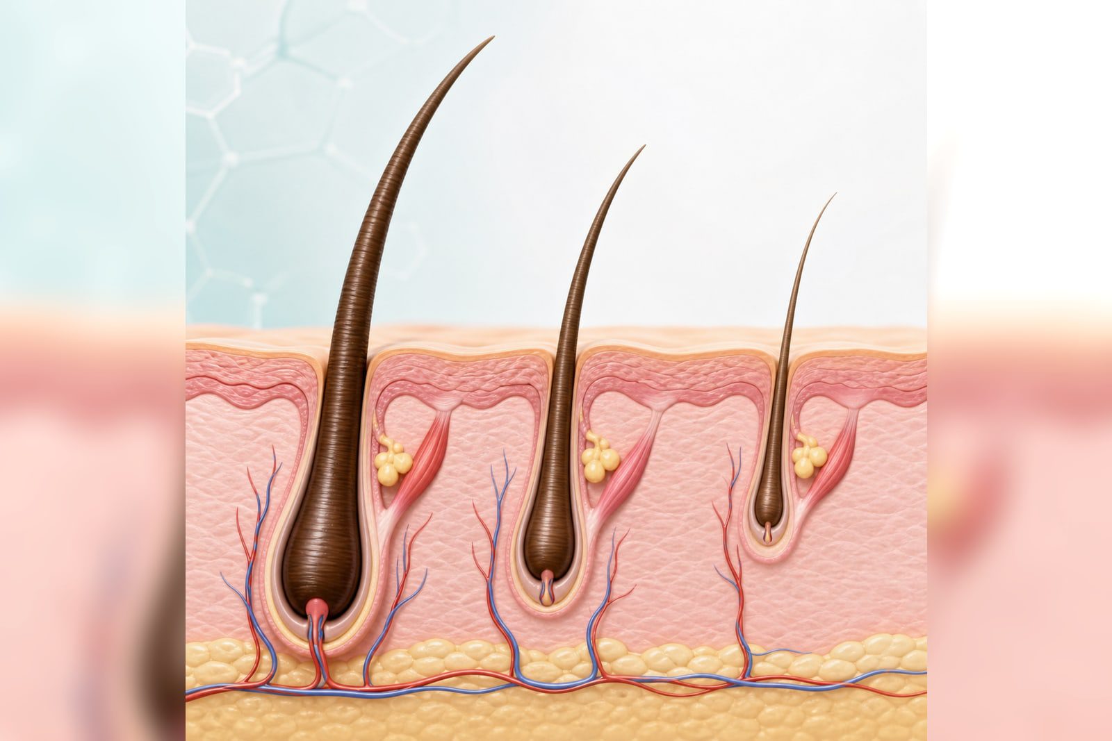
Losing a hair does not mean that follicle has disappeared. Hair is naturally released during its growth cycle; the important question is whether follicles are temporarily resting, gradually miniaturising, or being damaged by inflammation and scarring.
The amount seen in a shower or on a pillow cannot establish follicle function. Distribution, hair-shaft diameter, visible follicular openings and scalp redness, itch, pain or scale provide more useful clues.
Key points: A sudden diffuse increase often reflects a cycle shift and the follicles usually remain. Gradual thinning at the hairline, crown or part can reflect miniaturisation. Smooth shiny loss with fewer follicular openings, pain, burning or inflammation needs assessment for scarring alopecia. These cannot be diagnosed by appearance alone.
Does shedding mean dead follicles? Three different states
Temporary resting: the hair is shed but the follicle may remain
Fever, illness, surgery, childbirth, psychological stress, rapid weight change or nutritional disruption can move more follicles into a resting phase. Diffuse shedding appears later, while openings usually remain and new short hairs may be visible.
If a major stressor occurred in recent months, review the timeline and warning signs of stress-related shedding.
Gradual miniaturisation: new hairs become finer and shorter
Male or female pattern hair loss can progressively shorten the growth phase and convert terminal hairs into finer, shorter hairs. This is gradual miniaturisation rather than sudden follicle death.
Scarring damage: active inflammation needs early recognition
Some inflammatory scalp disorders can destroy follicles and replace them with scar tissue. A follicle already replaced by fibrosis is unlikely to produce hair again, so limiting ongoing inflammation is the priority.
What can the pattern of hair loss reveal?
Distribution and speed of change are more informative than a one-day hair count. Diffuse shedding, gradual fixed-pattern thinning, a sudden round patch and inflammatory scalp symptoms point to different assessments.
For receding temples or fixed crown thinning, see early signs and stages of male pattern hair loss; For a sudden defined patch, review alopecia areata features and assessment.
| Possible state | Typical distribution and appearance | Follicle clue | Suggested direction |
|---|---|---|---|
| Telogen effluvium | Sudden diffuse increase without a defined patch | Openings remain; short regrowth may appear | Review earlier triggers and track density |
| Pattern hair loss | Hairline, crown or part gradually thins | More shaft-diameter variation and fine hairs | Confirm pattern and miniaturisation |
| Alopecia areata | Sudden round or well-defined patch | Follicles remain; broken hairs may be present | Early dermatology assessment |
| Inflammatory scalp loss | Red, itchy, painful, scaly, pustular or crusted | Inflammation around follicles | Identify and treat scalp disease |
| Scarring alopecia | Smooth shiny area with fewer openings | Follicles may be replaced by fibrosis | Assess activity promptly |
How does trichoscopy assess follicle activity?
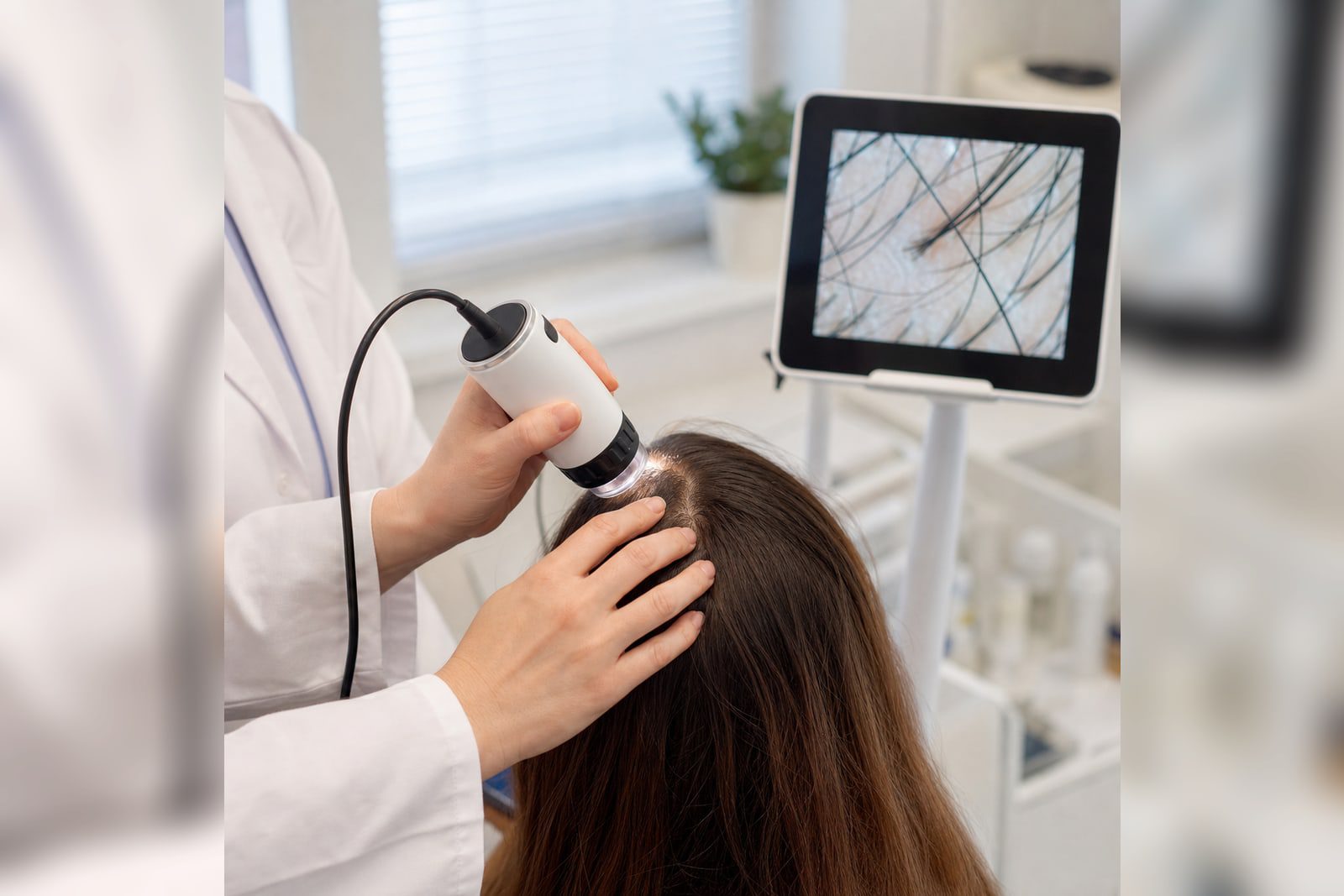
Trichoscopy is a non-invasive magnified examination of density, variation in shaft diameter, broken and new hairs, follicular openings, scale and perifollicular inflammation. It helps distinguish non-scarring from scarring loss.
Results are interpreted with the timeline, distribution, pull test, health, diet, weight change and family history. Blood tests are selected when clues justify them; biopsy is reserved mainly for suspected scarring loss or an unclear diagnosis.
Do fine short hairs always mean recovery?
They may be new growth or miniaturised hairs. Persistent diameter variation and falling density in one area differ from diffuse shedding followed by progressively longer, normal-calibre regrowth.
Can follicle miniaturisation improve?
“Follicle shrinkage” is not one diagnosis. A cycle shift calls for identifying the trigger and allowing time; pattern loss may require slowing miniaturisation and preserving existing hair; inflammation requires treatment of the scalp process.
Options depend on sex, age, pregnancy plans, health conditions, current treatment and diagnosis. The same topical product, oral approach or supplement should not be applied to everyone.
If male pattern loss is confirmed, compare oral and scalp-local treatment choices.
Which common approaches can mislead you?
Judging severity only by hair in the drain
Wash interval, hair length and grooming affect what you see. Standardised photographs and density trends over weeks or months are more useful.
Taking high-dose single nutrients without assessment
Deficiency can contribute to loss, but shedding alone does not prove deficiency. Testing and supplementation should follow the history and clinical assessment.
Using only hair-care products on a red or crusted scalp
Persistent redness, itch, greasy scale or pustules may signal scalp disease. Read about seborrhoeic dermatitis and other scalp disorders.
Which changes need prompt follicle assessment?
Brief mild shedding without density change can be documented. Arrange dermatology or hair-loss assessment if any of the following occurs:
- The hairline, crown or part keeps thinning and hairs become noticeably finer
- A sudden round, patchy or asymmetric area appears
- A bald area looks smooth and shiny with fewer follicular openings
- Persistent redness, itch, pain, burning, heavy scale, pustules, weeping or crusting
- Eyebrows, eyelashes or other body hair also fall
- Fatigue, marked weight change, cold intolerance, palpitations or menstrual change accompanies loss
view Ruianc Clinic’s hair-loss assessment. A consultation uses the timeline, pattern, trichoscopy and individual risks to identify the follicle state before deciding on follow-up or treatment.
Which sources support this article?
These general dermatology sources informed this article. Individual diagnosis and treatment still require clinical assessment.
- American Academy of Dermatology: Hair loss causes
- DermNet: Trichoscopy
- British Association of Dermatologists: Telogen effluvium
Not necessarily. Telogen effluvium, alopecia areata and some inflammatory conditions can increase shedding while follicles remain. Pattern, openings, shaft diameter and trichoscopy help distinguish the cause.
Miniaturisation produces progressively finer and shorter hairs. Scarring destruction can replace the follicle with fibrosis; the implications and management differ.
Appearance alone is not enough. Smooth shine with absent openings, redness, pain, burning or scale warrants assessment for scarring loss.
Either is possible. Regrowth lengthens over time; miniaturised hairs often remain fine and short in a fixed area. Serial photographs and trichoscopy help.
No. History, pattern and trichoscopy come first. Blood tests follow specific clues, while biopsy is mainly for suspected scarring or unresolved cases.
Early diagnosis of progressive pattern or inflammatory loss can preserve options and reduce further damage, but the cause must be established first.
Doctor’s note: follicles are not simply “alive” or “dead”
Shedding may reflect a cycle shift, gradual miniaturisation or inflammatory destruction. The clinician first checks distribution, follicular openings, shaft diameter and scalp symptoms before selecting follow-up or further tests.
This article is general health education, not an advertisement or recommendation for any specific medicine. Diagnosis, investigations and treatment require an in-person assessment by a qualified doctor.
Hair-loss medical team

Dr. Tseng